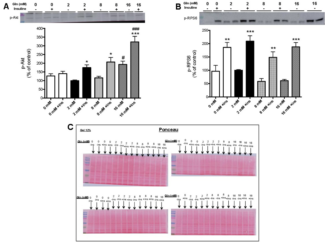
Fig. 6. Contents of phosphorylated Akt and RPS6 in C2C12 myotubes cultivated in various glutamine concentration conditions (no addition or 2, 8 or 16 mM glutamine) for 48 hours stimulated with insulin (100nM) in the last hour. (A) p-Akt and (B) p-RPS6 contents. (C) Ponceau S staining of nitrocellulose membranes after protein transfer from 12% polyacrylamide gels. The experimental groups are: (0 mM) no glutamine addition in the medium; 2 mM (reference condition), 8 mM or 16 mM L-glutamine in the medium. The findings are as the mean ± SEM of three independent experiments. Results were analyzed using one-way ANOVA and Tukey post-test. (*p<0.05; **p<0.01; ***p<0.001) compared with the respective group without insulin; (#p<0.001) compared with 2 mM glutamine; (###p<0.001) compared with 2 mM glutamine plus insulin. Outliers were excluded by applying Grubbs's test. The western blotting images of the results are in the Supplementary Fig. S2.